
Genetic disease mutations
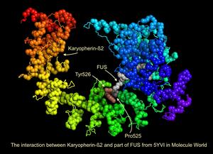
These structures show human proteins where missense mutations are associated with disease. You can view these structures, by downloading the collection and open it in the Molecule World iPad app.
Displaying 1 - 3 of 3
| Type | Structure Description | PDB ID | Download Structure | |
|---|---|---|---|---|
| An MW Collection | Image
|
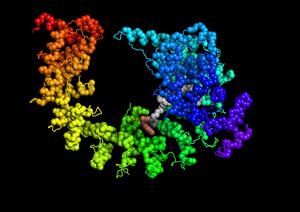
Download and open in the Molecule World iPad app to compare the normal and mutant structures. |
Document
Genetic_disease.mwc
(573.77 KB)
|
|
| PDB | Image
|
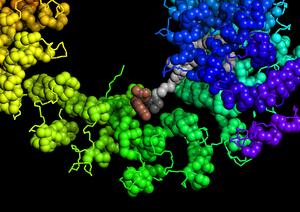
Karyopherin-beta2 bound to a portion of the normal FUS protein with proline 525. |
5YVI | Document
5yvi.pdb.gz
(291.15 KB)
|
| PDB | Image
|
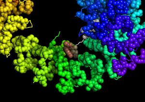
Karyopherin-Beta2 bound to a portion of the FUS protein, with a hypothetical model of a Pro525leu mutation. Note - this exact structure wouldn't exist because of steric hindrance. |
5YVI | Document
5yvi_P525L_mut.pdb.gz
(285.82 KB)
|